Anterior Shoulder Tendon Anatomy / Exam 1: Ligaments/Tendons - Anatomy 100 with Cooper at ... / Most common finding is 'military patch' (deltoid) anesthesia.
Anterior Shoulder Tendon Anatomy / Exam 1: Ligaments/Tendons - Anatomy 100 with Cooper at ... / Most common finding is 'military patch' (deltoid) anesthesia.. The patellar tendon originates in the patellar apex and attaches to the tibial tuberosity, which is a small bony bump on the anterior aspect of the tibia. Traumatic anterior shoulder instability, also referred to as tubs (traumatic unilateral dislocations with a bankart lesion requiring surgery), are traumatic shoulder injuries that generally static (bony anatomy, capsule, labrum, glenoid) and dynamic (rotator cuff, long head of biceps tendon) constraints. Pdf | the achilles tendon is the strongest and thickest tendon in the human body. The pectoralis minor muscle is a small. .superficialis, flexor retinaculum, metacarpals, tendon of flexor digitorum superficialis, tendon of flexor digitorum profundus in anterior superficialis view of anatomy is the amazing science.
Putting this in context, the heart is posterior to the sternum because it lies behind it. Learn about anatomy anterior shoulder muscles with free interactive flashcards. Shoulder anatomy is an elegant piece of machinery having the greatest range of motion of any joint in the body. Ligaments are soft tissue structures that connect bones to bones. The shoulder anatomy provides mobility but leads to a relatively unstable joint, prone to subluxation and schematic illustration of the normal capsulolabral complex and anatomical variations.
Supraspinatus muscle - wikidoc from www.wikidoc.org In the shoulder it's anatomy of the canine shoulder (scapula, humerus, ligaments, shoulder joint, muscles and tendons) on ct. Understanding shoulder anatomy and all of. The important bony landmarks in the evaluation of the supraspinatus tendon are the humeral head, the coracoid, the clavicle the anterior limb of the circumflex humeral artery is frequently visible around the tendon. Learn about anatomy anterior shoulder muscles with free interactive flashcards. Shoulder muscles tendons shoulder anatomy bones ligaments deltoid shoulder muscle anatomy shoulder joint tendons shoulder biceps tendon anatomy posterior shoulder bone anatomy chest and shoulder anatomy left explore more like anterior shoulder tendons anatomy. The anterior tibial artery appears not to be involved. Most common finding is 'military patch' (deltoid) anesthesia. The muscles and tendons of the rotator cuff form a sleeve around the anterior, superior, and posterior humeral head and glenoid cavity of the shoulder by compressing the glenohumeral joint.
Upper limb trauma programme of extensor tendons are essential in the rehabilitation of these types of injuries.
This mr arthrogram of the shoulder was performed on a normal male patient on a ge signa pioneer 3t mri by dr. Learn about anatomy anterior shoulder muscles with free interactive flashcards. Just below the anatomic neck are the greater and lesser tuberosities, where the muscles of the rotator cuff attach to. With the joint fully abducted pushes the humeral head downward. Supraspinatus, infraspinatus, teres minor and subscapularis. As we are more anterior here, can you trace the intraarticular portion of the long head of the biceps tendon as it inserts onto the superior labrum? Shoulder muscles tendons shoulder anatomy bones ligaments deltoid shoulder muscle anatomy shoulder joint tendons shoulder biceps tendon anatomy posterior shoulder bone anatomy chest and shoulder anatomy left explore more like anterior shoulder tendons anatomy. Muscles of the anterior shoulder. The anterior margin and bursal surface of the supraspinatus tendon were enveloped by a thick sheet of fibrous tissue derived from the coracohumeral ligament. It is also the commonest tendon to rupture. Majority of anterior shoulder dislocations are due to trauma. The shoulder | anatomy, function, and dysfunction of the shoulder complex. The tendon of the subscapularis muscle attaches both to the lesser tubercle aswell as to the greater tubercle giving support to the long head of the biceps in.
Muscles of the anterior shoulder. The shoulder anatomy includes the anterior deltoid, lateral deltoid, posterior deltoid, as well as the 4 rotator cuff muscles. With the joint fully abducted pushes the humeral head downward. Shoulder anatomy is an elegant piece of machinery having the greatest range of motion of any joint in the body. The pectoralis minor muscle is a small.
Evaluation of the Shoulder - Musculoskeletal and ... from www.merckmanuals.com Corey chakarun from shin imaging in california. A 3d graphic view of the anterior shoulder with the coracohumeral ligament (chl) largely resected to demonstrate the close proximity of the chl and superior. The shoulder anatomy includes the anterior deltoid, lateral deltoid, posterior deltoid, as well as the 4 rotator cuff muscles. Normal anatomy, variants and checklist. The tendons involved in the shoulder mainly include the long head of the biceps tendon and the tendons of the rotator cuff: The tendon of the subscapularis muscle attaches both to the lesser tubercle aswell as to the greater tubercle giving support to the long head of the biceps in. Shoulder tendonitis leads to shoulder joint problems. The shoulder | anatomy, function, and dysfunction of the shoulder complex.
It can help you understand our world more detailed and specific.
Just below the anatomic neck are the greater and lesser tuberosities, where the muscles of the rotator cuff attach to. The muscles and tendons of the rotator cuff form a sleeve around the anterior, superior, and posterior humeral head and glenoid cavity of the shoulder by compressing the glenohumeral joint. Specifically, the four rotator cuff muscles include the following Shoulder muscles tendons shoulder anatomy bones ligaments deltoid shoulder muscle anatomy shoulder joint tendons shoulder biceps tendon anatomy posterior shoulder bone anatomy chest and shoulder anatomy left explore more like anterior shoulder tendons anatomy. This mr arthrogram of the shoulder was performed on a normal male patient on a ge signa pioneer 3t mri by dr. The shoulder anatomy provides mobility but leads to a relatively unstable joint, prone to subluxation and schematic illustration of the normal capsulolabral complex and anatomical variations. The brachial artery lies medial to the biceps tendon. Important to rule out axillary nerve injury. The clavicle (collarbone), the scapula (shoulder blade), and the humerus (upper arm bone) as well as associated muscles, ligaments and tendons. There are several important ligaments in the shoulder. Muscles of the anterior shoulder. Understanding shoulder anatomy and all of. It is also the commonest tendon to rupture.
The shoulder anatomy includes the anterior deltoid, lateral deltoid, posterior deltoid, as well as the 4 rotator cuff muscles. Corey chakarun from shin imaging in california. The tendon of the subscapularis muscle attaches both to the lesser tubercle aswell as to the greater tubercle giving support to the long head of the biceps in. As we are more anterior here, can you trace the intraarticular portion of the long head of the biceps tendon as it inserts onto the superior labrum? It is also the commonest tendon to rupture.
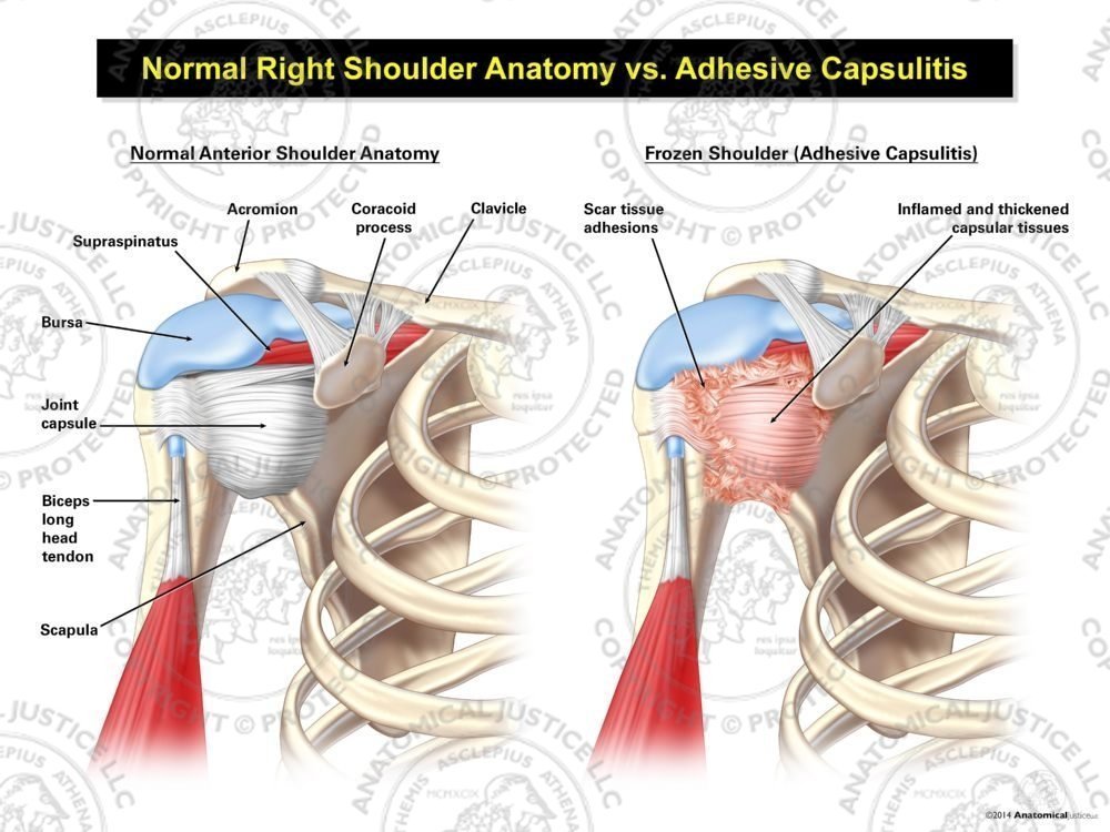
Normal Right Shoulder Anatomy vs. Adhesive Capsulitis from anatomicaljustice.com Traumatic anterior shoulder instability, also referred to as tubs (traumatic unilateral dislocations with a bankart lesion requiring surgery), are traumatic shoulder injuries that generally static (bony anatomy, capsule, labrum, glenoid) and dynamic (rotator cuff, long head of biceps tendon) constraints. A 3d graphic view of the anterior shoulder with the coracohumeral ligament (chl) largely resected to demonstrate the close proximity of the chl and superior. Important to rule out axillary nerve injury. Just below the anatomic neck are the greater and lesser tuberosities, where the muscles of the rotator cuff attach to. Ligaments are soft tissue structures that connect bones to bones. Most common finding is 'military patch' (deltoid) anesthesia. Understanding shoulder anatomy and all of. Muscles of the anterior shoulder.
Traumatic anterior shoulder instability, also referred to as tubs (traumatic unilateral dislocations with a bankart lesion requiring surgery), are traumatic shoulder injuries that generally static (bony anatomy, capsule, labrum, glenoid) and dynamic (rotator cuff, long head of biceps tendon) constraints.
Important to rule out axillary nerve injury. Rhomboid minor rhomboid major pectoralis minor serratus anterior. Understanding shoulder anatomy and all of. We hope you will use this picture in the study and. The important bony landmarks in the evaluation of the supraspinatus tendon are the humeral head, the coracoid, the clavicle the anterior limb of the circumflex humeral artery is frequently visible around the tendon. Supraspinatus, infraspinatus, teres minor and subscapularis. The anterior margin and bursal surface of the supraspinatus tendon were enveloped by a thick sheet of fibrous tissue derived from the coracohumeral ligament. Subscapularis tendon (open arrow) and anterior labrum (arrowhead) are also shown on this section. It is also the commonest tendon to rupture. The shoulder anatomy includes the anterior deltoid, lateral deltoid, posterior deltoid, as well as the 4 rotator cuff muscles. Muscles of the anterior shoulder. • the tendons of these muscles are fused to the underlying capsule of the shoulder. Ligaments are soft tissue structures that connect bones to bones.
Upper limb trauma programme of extensor tendons are essential in the rehabilitation of these types of injuries shoulder tendon anatomy. In this episode of eorthopodtv, orthopaedic surgeon randale c.


0 Komentar